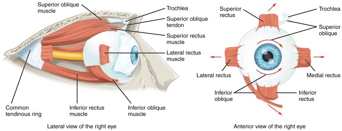
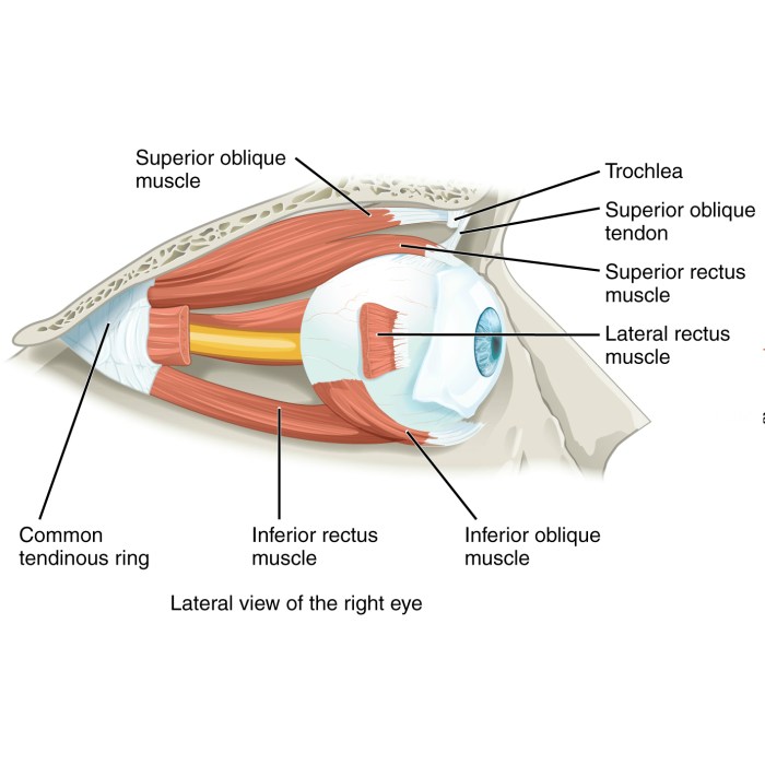
Label the extrinsic muscles of the eye – Labeling the extrinsic muscles of the eye is a fundamental aspect of understanding ocular anatomy and function. These muscles, innervated by cranial nerves, control the precise movements of the eye, enabling us to focus on objects, track moving targets, and maintain binocular vision.
This guide will delve into the location, actions, and innervation of these crucial muscles, providing a comprehensive overview for students, practitioners, and anyone seeking to enhance their knowledge of the visual system.
Extrinsic Muscles of the Eye: Overview

The extrinsic muscles of the eye are six muscles that control the movement of the eyeball. They are located outside the eye and are innervated by cranial nerves.
The extrinsic muscles of the eye are responsible for the following movements:
- Upward gaze
- Downward gaze
- Lateral gaze
- Medial gaze
- Intorsion
- Extorsion
The extrinsic muscles of the eye are innervated by the following cranial nerves:
- Oculomotor nerve (CN III)
- Trochlear nerve (CN IV)
- Abducens nerve (CN VI)
Rectus Muscles: Label The Extrinsic Muscles Of The Eye
The four rectus muscles are named according to their direction of action:
- Superior rectus muscle
- Inferior rectus muscle
- Lateral rectus muscle
- Medial rectus muscle
The rectus muscles are innervated by the oculomotor nerve (CN III), except for the lateral rectus muscle, which is innervated by the abducens nerve (CN VI).
| Rectus Muscle | Action | Innervation |
|---|---|---|
| Superior rectus muscle | Elevates the eye | Oculomotor nerve (CN III) |
| Inferior rectus muscle | Depresses the eye | Oculomotor nerve (CN III) |
| Lateral rectus muscle | Abducts the eye | Abducens nerve (CN VI) |
| Medial rectus muscle | Adducts the eye | Oculomotor nerve (CN III) |
Oblique Muscles

The two oblique muscles are named according to their direction of action:
- Superior oblique muscle
- Inferior oblique muscle
The superior oblique muscle is innervated by the trochlear nerve (CN IV), while the inferior oblique muscle is innervated by the oculomotor nerve (CN III).
- Superior oblique muscle:Elevates, intorts, and abducts the eye
- Inferior oblique muscle:Elevates, extorts, and abducts the eye
Innervation of the Extrinsic Muscles of the Eye

The extrinsic muscles of the eye are innervated by the following cranial nerves:
- Oculomotor nerve (CN III): Superior rectus, inferior rectus, medial rectus, and inferior oblique muscles
- Trochlear nerve (CN IV): Superior oblique muscle
- Abducens nerve (CN VI): Lateral rectus muscle
| Cranial Nerve | Extrinsic Muscles Innervated |
|---|---|
| Oculomotor nerve (CN III) | Superior rectus, inferior rectus, medial rectus, and inferior oblique muscles |
| Trochlear nerve (CN IV) | Superior oblique muscle |
| Abducens nerve (CN VI) | Lateral rectus muscle |
Clinical Significance
The extrinsic muscles of the eye are essential for normal vision. Dysfunction of these muscles can lead to a variety of conditions, including:
- Strabismus (misalignment of the eyes)
- Ptosis (drooping of the eyelid)
Strabismus is a condition in which the eyes are misaligned. This can be caused by a variety of factors, including weakness or paralysis of the extrinsic muscles of the eye.
Ptosis is a condition in which the eyelid droops. This can be caused by weakness or paralysis of the levator palpebrae superioris muscle, which is innervated by the oculomotor nerve (CN III).
Clarifying Questions
What are the four rectus muscles of the eye?
The four rectus muscles are the superior rectus, inferior rectus, medial rectus, and lateral rectus.
Which cranial nerve innervates the superior oblique muscle?
The superior oblique muscle is innervated by the trochlear nerve (cranial nerve IV).
What is the function of the lateral rectus muscle?
The lateral rectus muscle abducts (moves laterally) the eye.